Core Facilities, Centers and Institutes
Increasingly, these sophisticated facilities are becoming critical state, national and international assets, supporting projects and programs of student and public benefit, including federally funded national centers of excellence.
RESEARCH OFFICE BASE CORE FACILITIES:
Core facilities reporting to Research Office 
COLLEGE BASED CORE FACILITIES:
CANR: College of Agriculture and Natural Resources
CAS: College of Arts and Sciences
CHS: College of Health Sciences
 Advanced Materials Characterization Lab Core Facility
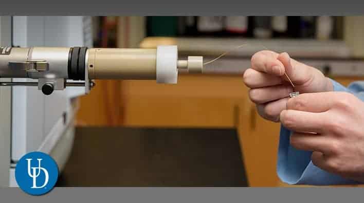
Advanced Materials Characterization Lab Core Facility
DIRECTOR: Gerald Poirier
ADDRESS: 221 Academy St, Newark, DE 19716
LOCATED IN: Interdisciplinary Science and Engineering Laboratory
Core Facility OVERVIEW: The University of Delaware’s Advanced Materials Characterization Lab is centrally located on the ground floor of the Interdisciplinary Science and Engineering building. This provides the opportunity and setting for the exchange of novel ideas and science for the next generation. Located in the same building are the multidisciplinary classrooms for undergraduate education continuing the theme of the multidisciplinary approach.
EQUIPMENT:
- Phillips Xpert Powder X-ray Diffractometer
- Rigaku Miniflex Powder X-ray Diffractometer
- Rigaku Ultima 4 Multipurpose X-ray Diffractometer
- Bruker D8 Multipurpose X-ray Diffractometer
- Phillip SAXS small angle Diffractometer
- T/A Instruments DSC,DTA,TGA,DMA,DTC
- Metrohm IC Pro Chromatograph
- Agilent HPLC 1260 /MS 6120
- Agilent Cary UV Vis Spectrometer
- Kaiser 785nm Raman Spectrometer
- Wyatt Dynamic Light Scattering
- Beckman Coulter LS13 320 Particle Analyzer
- Analsys NanoIR2
- Micromeritics Porosity Analyzer
- Agilent ICP/MS 7500
- Agilent AES 4100 Mass Spec.
SERVICES:
Provide short courses on all instrumentation on a regular basis.
 Bioinformatics Data Science Core Facility

Bioinformatics Data Science Core Facility
DIRECTOR: Shawn W. Polson, Ph.D.
ADDRESS: Delaware Biotechnology Institute, 15 Innovation Way, Suite 205, Newark, DE 19711
CONTACT: CBCB Core Facility
Core Facility and Center OVERVIEW: Providing scientific expertise and core infrastructure support in Bioinformatics and Computational Biology for the Delaware research and education community. Available resources include bioinformatics services, consulting, training and access to computational infrastructure and software.
 Center for Biomedical and Brain Imaging Core Facility
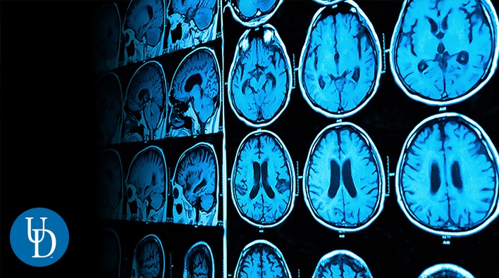
Center for Biomedical and Brain Imaging Core Facility
DIRECTOR: Keith Schneider, Ph.D.
ADDRESS: 77 E. Delaware Ave., Newark, DE 19716
CONTACT: Trevor Wigal, Manager, CBBI
(Sq. ft.: ~ 11,600)
Center, Core Facility and Center OVERVIEW: Serving researchers campus-wide, statewide and throughout the region, the CBBI will advance research on psychopathology, cancer, stroke, cerebral palsy, osteoporosis and other diseases and disorders.
 Center for Data-Intensive & Computational Science
Center for Data-Intensive & Computational Science
DIRECTOR: Rudi Eigenmann, Ph.D.
ADDRESS: Suite 147, 590 Avenue 1743, Newark, DE 19713
CONTACT: Data Science Institute
LOCATED IN: Ammon Pinizzotto Biopharmaceutical Innovation Center
Core Facility and Center OVERVIEW: The Data Intensive & Computational Science (DiCoS) Core supports interdisciplinary research collaborations to achieve competitiveness for funding opportunities, in particular major federal grants involving large, multidisciplinary teams.
DiCoS aims to advance both the research frontiers and the underlying infrastructure at the nexus of computational and data science, enabled by high-performance computing and big data. The integration of computational science, and data-intensive research is a central mission.
As a UD Core Facility, DiCoS develops and guides the use of shared resources and scientific expertise to support data science and computational research across campus. DiCoS identifies the needs of the research and education community, providing scientific expertise and infrastructure support. It leverages available resources and capabilities at UD and collaborates closely with UD Information Technologies and the Research Office.
SERVICES:
- Computational Resources
- Storage Resources
- Datasets
- Expert Assistance for Developing Computational and Data-intensive Applications
- Strategic Documents of Funding Agencies
- Training
 Center for Human Research Coordination Core Facility
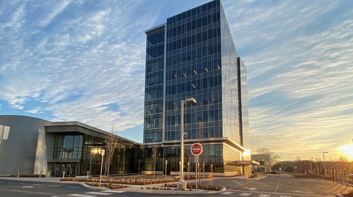
Center for Human Research Coordination Core Facility
LEADERSHIP: Karin Silbernagel, Ph.D., PT, ATC
ADDRESS: 100 Discovery Blvd. 10th Floor, Newark, DE 19713
CONTACT: Marlo Goss, Assistant Director
LOCATED IN: STAR Tower
Core Facility OVERVIEW: The Center for Human Research Coordination (CHRC) is a university-wide Core facility, established in July 2021, the Center supports UD Researchers focused on human subject research.
CHRC is focused on supporting researchers with participant recruitment, both on a small and large scale. The center is aimed at ensuring participant recruitment reflects the population and community we serve, and improving awareness of University of Delaware research across Delaware and neighboring Maryland and Pennsylvania communities. CHRC coordinates research subject logistics such as screening, scheduling, transportation, and reimbursement. CHRC also provides the university community with data management education, database set-up, repository, maintenance, hardening and security. CHRC supports REDCap across UD and assists researchers in building and maintaining participant registries.
SERVICES:
- Participate in Research Studies
- Join the Community Participant Registry
- Researchers Access
- Study Coordination
- Recruitment and more
 DBI BioImaging Center Core Facility

DBI BioImaging Center Core Facility
DIRECTOR: Jeffrey L. Caplan, Ph.D.
ADDRESS: Avenue 1743, Newark, DE 19713
LOCATED IN: Ammon Pinizzotto Biopharmaceutical Innovation Center, Suite 141
Core Facility and Center OVERVIEW: The BioImaging Center is a multi-user microscopy facility containing state-of-the-art electron, confocal and light microscopes. The center is open to all academic researchers on a fee-for-service basis. Outside industrial users are accommodated when scheduling permits.
EQUIPMENT:
We host a range of microscopes and sample preparation equipment to meet research demands.
Microscopes:
- Zeiss LIBRA 120 transmission electron microscope (TEM)
- Hitachi S4700 field emission scanning electron microscope (FE-SEM)
- Zeiss LSM780 spectral and high sensitivity confocal microscope
- Zeiss LSM510 NLO multi-photon confocal microscope
- Zeiss LSM510 DUO spectral and high-speed confocal microscope
- PECON live cell incubator for the inverted confocal microscopes
- INSTEC thermoelectric heating and cooling for confocal microscopes
- Zeiss ELRYA PS1 super resolution microscope
- Custom-built total internal reflection fluorescence microscope (TIRFM)
- TIRFM for single molecule experiments
- Zeiss M2BIO stereo dissecting light microscope
- Zeiss Axioplan2 upright light microscope
- Zeiss PALM COMBI laser capture microdissection microcope (LCM)
- Veeco Nanoscope IIIA atomic force microscope (AFM)
- Bruker Bioscope Catalyst AFM with integrated light microscopy
Sample Preparation equipment:
- Leica EM AFS automated freeze substitution system
- Leica EM PACT high-pressure freezing system
- Leica EM CPC plunge freezer
- Leica EM IGL automated immunogold labeler
- Reichert-Jung Ultracut E microtome for ultrathin sectioning (two)
- PELCO Biowave 34700 microwave tissue processor
- PELCO easiGlow Glow Discharge system
- Leica EM KMR2 glass knife maker
- EMCorp Microcut 1200 Vibratome
- Denton Bench Top Turbo III for carbon or gold/palladium coating
- Autosamdri-815B critical point dryer
Analysis tools:
- Dell T7500 Workstation for super resolution analysis
- Dell T5500 Workstation for visualization software
- VSG AMIRA 5.4 analysis and visualization software
- Perkin Elmer Volocity 6.2 analysis and visualization software
- SVI Huygens Pro deconvolution software
SERVICES:
We provide full-service sample preparation, data acquisition and image processing for all imaging technologies upon request. Experienced staff members can provide training on equipment, sample preparation techniques, and image analysis.
 DNA Sequencing and Genotyping Center
DNA Sequencing and Genotyping Center
DIRECTOR: Bruce Kingham, Ph.D.
ADDRESS: 590 Avenue 1743, Suite 146, Newark, DE 19711
CONTACT: UD Sequencing & Genotyping Center
LOCATED IN: Ammon-Pinizzotto Biopharmaceutical Innovation Center
Core Facility and Center OVERVIEW: The University of Delaware Sequencing and Genotyping Center (SGC) supports genomics research through our established expertise with state-of-the-art genomics technologies. Our core center is located in the Delaware Biotechnology Institute, which is an interdisciplinary research unit at the University of Delaware. The SGC provides assistance with experimental design, user training, sample and data analysis. Instrumentation housed in the SGC supports high-throughput, single-molecule, and Sanger sequencing technologies, as well as qualitative and quantitative analysis of nucleic acids.
EQUIPMENT:
- llumina HiSeq 2500
- Pacific Biosciences RSII Single-Molecule Sequence
- ABI Prism 3130XL Genetic Analyzer
- ABI 7500 Fast Real-Time PCR System
- Pippin Blue DNA Size Selection System
- Covaris S2 Adaptive Focused Acoustic Disruptor with CryoPrep
- Agilent 2100 Bioanalyzer
- Qubit Fluorometer
- NanoDrop Technologies ND-1000 UV-Vis Spectrophotometer
- MJ Research Tetrad Thermocycler
- ABI Veriti and GeneAmp PCR System 9700 Thermocycler
- Eppendorf Centrifuge 5804R
SERVICES:
- DNA sequencing (ABI BigDye Sanger sequencing and Illumina SBS next-generation sequencing)
- Genotyping (fragment analysis microsatellite analysis, STR analysis)
- PCR, qPCR, DNA preparation and analysis services (including extraction, purification, quantitation and quality scoring, fragmentation, amplification, plasmid construction and preparation).
- Access to pyrosequencing and microarray instrumentation
 Keck Center for Advanced Microscopy and Microanalysis Core Facility (a subdivision of the Advanced Materials Characterization Lab)
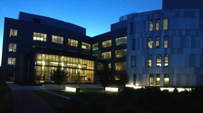
Keck Center for Advanced Microscopy and Microanalysis Core Facility (a subdivision of the Advanced Materials Characterization Lab)
DIRECTOR: Gerald Poirier
ADDRESS: 250V Harker Interdisciplinary Science and Engineering Laboratory, Newark, DE 19716
LOCATED IN: Harker Interdisciplinary Science and Engineering (ISE) Laboratory
Core Facility OVERVIEW: The W.M. Keck Center for Advanced Microscopy and Microanalysis (Keck CAMM) is a subdivision of the Advanced Materials Characterization Lab and has been established since 2001 through generous grants from the W. M. Keck Foundation, the National Science Foundation and funds from the University of Delaware. This facility contributes to scientific capabilities by enabling students, faculty and other researchers in the University and from regional institutions and facilities to use state-of-the-art equipment for research and education.
It houses two 200 kV field emission transmission electron microscopes, Talos F200C and JEM-2010F, a LaB6 300kV TEM JEM-3010, an FEI 120kV Tecnai G2 12 Twin transmission electron microscope, two scanning electron microscopes (JSM-7400F and AURIGATM 60 CrossBeamTM with the AURIGATM 60 being a FIB-SEM dual beam instrument), and two scanning probe microscopes (Multimode NanoScope V and Dimension 3100 V).
EQUIPMENT:
TEM
- Equipment 1 Talos F200C (200kV FE-TEM)
- Equipment 2 JEM-2010F (200kV FE-TEM)
- Equipment 3 JEM-3010 (300kV TEM)
- Equipment 4 Tecnai G2 12 (120kV TEM)
SEM
- Equipment 5 Auriga 60 CrossBeam (FIB/FE-SEM)
- Equipment 6 JSM-7400F (FE-SEM)
AFM
- Equipment 7 Dimension-3100 V SPM
- Equipment 8 MultiMode NanoScope V SPM
SERVICES:
The laboratory staff and associated faculty members are knowledgeable in SEM, TEM and SPM and are experts in advanced microscopy of a wide range of materials. We work with users on campus and other organizations in many areas including research, research training, and consultation.
 Materials Growth Facility Core
Materials Growth Facility Core
DIRECTOR: Joshua M. O. Zide, Ph.D.
ADDRESS: 201 DuPont Hall, 127 The Green, Newark, DE 19716
CONTACT: Materials Growth Facility
LOCATED IN: Harker Interdisciplinary Science and Engineering (ISE) Laboratory
Core Facility OVERVIEW: The University of Delaware Materials Growth Facility (MGF) primary objective is to provide the infrastructure, equipment, and staff support necessary to enable existing faculty, new faculty, and academic and corporate partners to undertake competitive research and development in the growing number of materials, science, and engineering fields.
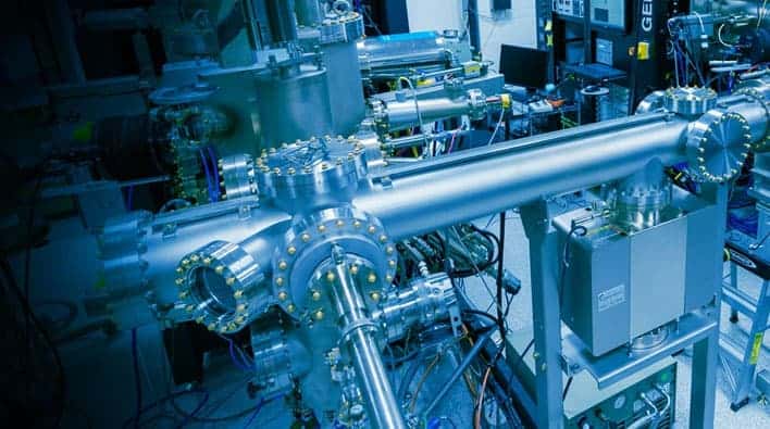
The University of Delaware (UD) Materials Growth Facility (MGF) offers III-V and topological insulator (TI) growth of epitaxial semiconductor films.
These growths are performed on a dual-chamber GENxplor molecular beam epitaxy (MBE) system. Our staff offers full-service material calibration and growth, as well as training to perform MBE deposition.
The MGF is integrated into the Delaware Institute for Materials Research (DIMR), providing seamless materials growth, materials characterization, electron microscopy, and nanofabrication capabilities.
EQUIPMENT:
III-V MBE (APOLLO) FEATURES:
- Standard III-V source materials (gallium, indium, aluminum, arsenic, antimony)
- Electronic doping sources (beryllium, tellurium, high-flux silicon)
- Bismuth and two rare earth sources (erbium, terbium)
- BandiT band-edge thermometry and thermocouple feedback for precise control of substrate temperature
- Atomic hydrogen beam cleaning
- Substrate heating up to 1200 °C (sample rotation up to 60 rpm)
- Sample size: small pieces to 3″ wafers
- In-situ monitoring: pyrometry, RHEED, RGA (to 100 AMU), chamber and BFM ionization gauges
- Load lock sample out-gassing (200 °C)
- Recipe driven
CHALCOGENIDE MBE (ARTEMIS) FEATURES:
- Topological insulators source materials (bismuth, indium, antimony, selenium, tellurium)
- Electron beam evaporator for high-temperature materials (tungsten, molybdenum, tantalum, niobium, zirconium)
- Low-flux gallium capping source
- Substrate heating up to 1200 °C (sample rotation up to 60 rpm)
- Sample size: small pieces to 3″ wafers
- In-situ monitoring: RHEED, RGA (to 100 AMU), chamber and BFM
- ionization gauges
- Load lock sample out-gassing (200 °C)
- Recipe driven
UHV MAGNETIC SPUTTERING SYSTEM (HEPHAESTUS) WILL FEATURE:
- A wide range of source targets (metallic, dielectric, and precious metal)
- Three high-purity gas controllers (argon, nitrogen, oxygen)
- Six magnetrons (three with high strength fields for magnetic target materials)
- Off-axis capability for one magnetron (for deposition of MgO based magnetic tunnel junctions)
- High temperature substrate heater
SERVICES:
CALIBRATIONS
The following calibrations are standard. Calibrations outside these standard calibrations may be the responsibility of the group needing them, warrant consideration as a collaborator, or eventually be included as a standard calibration.
III-V MBE
- GaAs, AlAs, and InAs growth rates
- GaSb and AlSb growth rates
- InGaAs lattice-matching (to InP) and growth rate
- InAlAs lattice-matching (to InP) and growth rate
- Silicon and beryllium doping in GaAs (generally convertible to other materials)
- Tellurium doping in GaSb
Chalcogenide MBE
- Bi2Se3 growth rate
- Bi2Te3 growth rate
- In2Se3 growth rate
Growths utilizing our wide range of additional Capabilities can be calibrated to specific user needs or performed on a “best effort” basis.
For additional details, see our MGF Procedures or contact us (below).
WAFERS
The MGF stocks several wafer types which are available to users at cost.
- 2” GaAs (001-oriented, semi-insulating)
- 2” GaAs (001-oriented, n-type)
- 2” InP (001-oriented, semi-insulating)
- 2” InP (001-oriented, n-type)
- 1cm x 1cm c-plane sapphire
- Other wafers may be used with approval
CHARACTERIZATION
The Epitaxy Engineer can perform sample characterization, using many of the facilities available at UD. When staff-assisted time that does not involve growth on an MBE is required, user fees are set on an hourly basis.
 Nanofabrication Facility
Nanofabrication Facility
DIRECTOR: Iulian Codreanu
ADDRESS: 221 Academy St., Newark, DE 19716
CONTACT: Iulian Codreanu
(Sq. ft.: ~ 8,500)
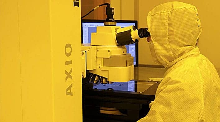
Core Facility OVERVIEW: The UD Nanofabrication Facility (UDNF) will enable researchers in academia, industry and government to create devices smaller than a human hair, supporting scientific advances in fields ranging from medical diagnostics to environmental sensing to solar energy harvesting. Explore our website to learn about our facility and all that we offer.
EQUIPMENT:
The UD Nanofabrication Facility (UDNF) has world-class capabilities in the areas of lithography, deposition, etch, thermal processing, characterization, and device packaging. Below is a comprehensive list of our equipment and fees.
Film Casting
• E-beam SBU1, • E-beam SBU2, • Photo SBU1, • Photo SBU@, • SU-8 SBU
Lithography
• E-beam Writer, • Laser Writer, • Mask Aligner
Deposition
• ALD, • Evaporator 1, • Evaporator 2, • PECVD, • PLD, • Sputterer
And much more. For a full listing of our equipment offerings click here.
SERVICES:
UDNF will consider performing fabrication work on behalf of others. The service fee is $50/hr for UD users, $75/hr for external academic users, and $150/hr for corporate users. Please contact Iulian Codreanu for details.
UD has many additional resources that may be of interest to potential facility users. The links below will take you to additional information on other user facilities on campus. Keck Center for Advanced Microscopy & Microanalysis, Advanced Materials Characterization Laboratory, Materials Growth Facility, Delaware Biotechnology Institute.
Our Services include: Film Casting, Lithography, Deposition, Dry Etch, Thermal Processing, Metrology, Packaging and more.
 Office of Laboratory Animal Medicine
Office of Laboratory Animal Medicine
LEADERSHIP: Eric Hutchinson, D.V.M.
CONTACT: Julie, pugsley@udel.edu
Core Facility OVERVIEW: The University of Delaware is committed to the proper care and humane treatment of all animals used in research and teaching at this institution. The IACUC will review, and if warranted, investigate all allegations, whether made by the public or an employee of this institution. Federal laws and regulations prohibit discrimination or reprisal for reporting any animal welfare concerns. Reports about animal welfare or non-compliance will be handled confidentially, if requested.
 Pearson Hall MakerSpace
Pearson Hall MakerSpace
ADDRESS: 125 Academy Street, Newark, DE 19716
CONTACT: Makerspace
LOCATED IN: Pearson Hall
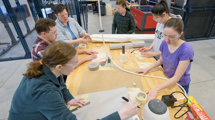
Core Facility OVERVIEW: The MakerSpace is an interdisciplinary design and fabrication studio, focused on student empowerment and collaboration. This creative space is equipped with a robust array of processes which compliment and provide depth to existing making capabilities on campus in order to support education, research, and personal growth. All students have access to our resources including the necessary training and design consultation to help them turn their ideas into action. This is everyone’s sandbox.
EQUIPMENT:
- Ultimaker S5 FDM 3D Printers (8)
- MarkForged X7 CFF 3D Printer
- Universal ILS12.75 Laser Cutter
- Universal PLS6.75 Laser Cutter
- Omax Protomax Water Jet
- Thermwood Multipurpose 45 CNC Router
- Tormach M1100MX CNC Mill
- Safety Speed H5 Panel Saw
- SawStop Industrial 5HP Table Saw
- Omga 1P 350F Miter Saw
- Felder FB510 Band Saw (2)
- Nova Voyager Drill Press
- Powermatic 31A Combination Sander
- Spindle Sander Jet JOSS-S
- Juki DDL-5550N Industrial Sewing Machine (2)
- Epson SureColor P9000 Standard Edition Printer
SERVICES:
- Use of the Pearson Hall MakerSpace resources is by reservation. Walk-in use is permitted provided it does not interfere with previously scheduled reservations or scheduled events.
- Your reservation will not be honored if you have not fully completed the required MakerSpace training and are not authorized on that equipment.
- MakerSpace Reservation
CANR: Comparative Pathology Laboratory Core Facility
CANR: Comparative Pathology Laboratory Core Facility
DIRECTOR: Erin Brannick, Ph.D.
ADDRESS: 15 Innovation Way, Newark, DE 19711
LOCATED IN: Delaware Biotechnology Institute
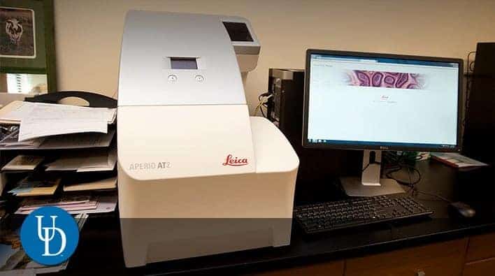
Core Facility OVERVIEW: The Comparative Pathology Laboratory provides histology and pathology support for animal diagnostic laboratories of Delaware and Maryland, researchers at the University of Delaware and its affiliates (DBI, etc.), industry and government partners of the university, and external researchers. We specialize in animal tissues and studies related to animal disease or animal models of human disease. However, we also routinely accept plant specimens for tissue processing. We strive to provide clients with customized high quality specimen preparations and thorough pathology reports to meet their individual diagnostic and research needs.
EQUIPMENT:
Leica Processor, Embedding Station, Microtome, Sakura Autostainer, Sakura Autostainer
SERVICES:
Histology Services: Tissue preparation including trimming, processing, paraffin embedding and microtomy, automated H&E staining, manual special staining upon request (i.e. Gram, Masson’s trichrome, etc.)
Consultation and equipment training upon request:
Pathology Services: Microscopic analysis of tissues for diagnostic cases and research studies, consultation on experimental design, necropsy, and specimen collection and preparation, digital photography of gross or histologic specimens.
CANR: Fischer Greenhouse Complex and Plant Growth Facilities Core Facility
CANR: Fischer Greenhouse Complex and Plant Growth Facilities Core Facility
LEADERSHIP: William Bartz
LEADERSHIP: Rodney Dempsey
ADDRESS: 531 South College Ave., Newark, DE 19716
LOCATED IN: Fischer Greenhouse Complex (Sq. ft.: ~ 33,600)
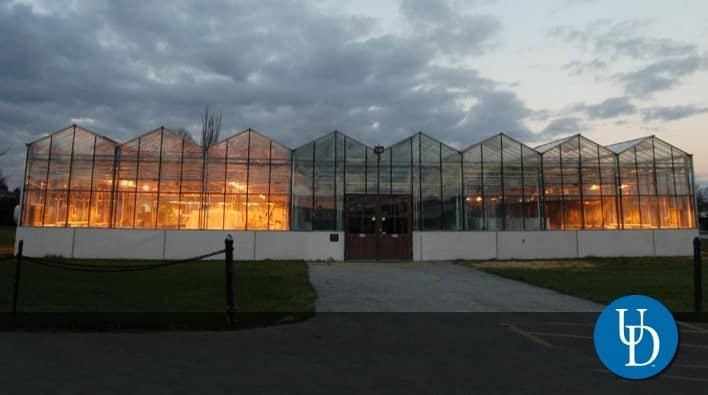
Core Facility OVERVIEW: A state-of-the-art plant growth facility in the University of Delaware College of Agriculture and Natural Resources, the Fischer Greenhouse Complex is a professionally-managed suite of growth chambers and glass house facilities serving the research and education community.
The Fischer Greenhouse Complex provides the primary greenhouse space available to faculty, professionals, staff and students, and is dedicated to the acquisition and dissemination of knowledge through research, teaching and outreach activities, extension demonstrations, departmental functions, and sponsored student organizations.
EQUIPMENT:
The 13,800 ft2 Fischer Greenhouse Complex and the Growth Chamber Facilities offers:
- Glass houses (17,000 ft2) with headhouse (5,000 ft2)
- Growth chambers (2,300 ft2 ) and growth room (500 ft2)
- Commercial Seed Dryer and Seed Storage Unit
SERVICES:
- Cold Storage for Plant Germplasm
CAS: High Throughput Experimentation Center
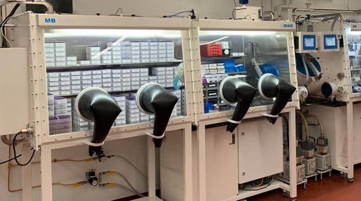
CAS: High Throughput Experimentation Center
LEADERSHIP: Jessica Sampson, Ph.D.
ADDRESS: 124 Lammont du Pont Lab, Newark, DE 19716
CONTACT: HTE Center
LOCATED IN: Lammont du Pont Laboratory
Core Facility OVERVIEW: The goals of the High Throughput Experimentation (HTE) Center at the University of Delaware are to enable high throughput chemical experimentation, including compound library synthesis, reaction development, and collection of data sets for statistical model development. We seek to do to this through the use of our extensive library of chiral and achiral ligands, automated and manual tools for reaction set-up, and analytical instrumentation for detection of products with a range of molecular weights, stabilities, and polarities. Initially funded by a UNIDEL grant received by Dr. Don Watson and Dr. Mary Watson, we are currently in the pilot phase for this lab and actively working to recruit new users and implement a charging model. Please feel free to reach out if you have any questions about using or collaborating with our facility.
SERVICES OFFERED
- Design, running, and analysis of 24- and 96-well plates, including for:
– Ligand screening - – Reaction optimization
– Library synthesis - LC-MS, SFC-MS, and GC-FID method development
- LC-MS, SFC-MS, GC-FID, and GC-MS analysis
- Training on facility instrumentation and on running HTE plates should be requested through iLab.
CAS: Mass Spectrometry Core Facility

CAS: Mass Spectrometry Core Facility
DIRECTOR: Papa Nii Asare-Okai, Ph.D.
ADDRESS: 110 Lammot duPont Laboratory, Newark, DE 19716
LOCATED IN: Lammot duPont Laboratory
Core Facility OVERVIEW: The core facility is grouped into three areas; the first laboratory houses the high performance instruments for more detailed and precise analyses, a second lab houses three open-access, user-friendly instruments that are available for researchers to use 24/7, and finally, a high-performance MALDI instrument is located in Murray V. Johnston’s Lab.
EQUIPMENT:
High-Performance Instrumentation Lab (Room 122, Lammot duPont Lab), Protein Sequencing, Micromass model Ultima Q Tof (Quadrupole-Time of Flight tandem mass spectrometer): This is an LC-MS-MS instrument primarily used to identify proteins by sequencing the products of a digested protein. Product molecular ions and subsequent collisional activation to generate fragments are automatic. Software generated or de novo peptide sequencing can be compared to protein databases for identification.
Protein Digester- Bruker Daltonics model Proteineer DP: This automated digest and prep station performs protein digests. Digest products can be automatically spotted to a MALDI sample plate or captured in MTP format for LC-MSMS analyses.
Open Access Instrumentation Lab (Room 108, Lammot duPont Lab)
GC-MS (Gas Chromatography-Mass Spectrometry): Agilent models 6850 GC and 5973 MS with autosampler. Used primarily for separation of small organic mixtures, incorporates a NIST library to aid identification.
LC-MS (Liquid Chromatography-Mass Spectrometry): Thermo-Finnigan model LCQ using Electrospray Ionisation (ESI) with an integrated autosampler. Used for biological samples and polar organics. The instrument can be switched between column mode for mixture separations and loop injections for quick analyses of purer samples.
MALDI (Matrix Assisted Laser Desorption Ionisation) Bruker Daltonics model Omniflex: Used primarily for biological samples to determine molecular weight and purity.
Murray V. Johnston Lab (Room 125, Lammot duPont Lab)
MALDI- Bruker Daltonics model Biflex MALDI mass spectrometer: Used for high-resolution and accurate mass measurements; automated protein identification via peptide mass maps.
SERVICES:
High-resolution accurate mass measurement (HRMS) of organic and organometallic compounds using Waters GCT Premier equipped with Electron Impact (EI), Chemical Ionization (CI), Desorption Chemical Ionization (DCI), Field Ionization (FI), Field Desorption (FD) and Liquid Injection Field Desorption Ionization (LIFDI).
LC/MS and LC/MS/MS using Shimadzu LCMS 2020, Waters Q-TOF and Thermo Q-Exactive Orbitrap with Electrospray Ionization (ESI) and Atmospheric Pressure Chemical Ionization (APCI).
GC-MS (low-resolution EI only) using the Agilent 5973 system.
MALDI analysis of proteins, oligonucleotides, nanoparticles, synthetic polymers and macromolecules.
Mass directed prep purification is also available.
CAS: NMR Laboratory Core Facility
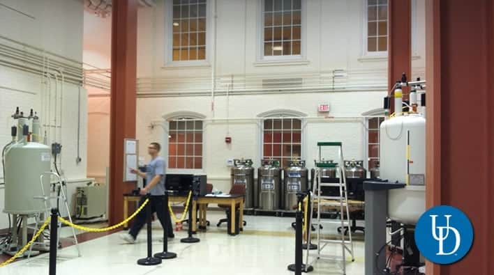
CAS: NMR Laboratory Core Facility
DIRECTOR: Steve Bai, Ph.D.
ADDRESS: 015 Brown Laboratory, Newark, DE 19716
LOCATED IN: Brown Laboratory (Sq. ft.: ~ 4,650)
Core Facility OVERVIEW: The Nuclear Magnetic Resonance (NMR) Facility currently has six NMR spectrometers. The NMR facilities are located in the basement floor of the North Wing of Brown Laboratories (BRL). The 600 MHz instrument is housed in 015 BRL and the remaining spectrometers are located across the hallway in three adjacent rooms (011, 011A and 021 BRL).
EQUIPMENT:
Two solution 400 MHz NMR spectrometers, one with an auto-sampler and a cryogenic QNP probe and another with multinuclear capabilities, are available for routine proton and multinuclear NMR analysis of organic and inorganic materials. Also: two solution 600 MHz spectrometers, one with a triple-resonance cryogenic probe for biomolecular samples and another with an auto-sampler and enhanced 19F capabilities for expanded NMR applications such as high-resolution magic angle spinning (HRMAS) for semi-solid materials.
The 500 MHz solid-state NMR spectrometer is currently equipped with a 3.2 mm triple-resonance (1H, 13C and 15N) probe for biosolids and other organic solid material. The 850 MHz spectrometer is hybridized for both solution and solid-state NMR measurements. With the ultra-high magnetic field and a large collection of solution and solid-state NMR probes, the 850 MHz NMR spectrometer covers a broad range of applications from inorganic material, synthetic organic polymeric materials to structure and dynamic studies in structural biology.
In addition to the departmental NMR instrument, four NMR spectrometers with frequencies ranging from 200 to 600 MHz are used and maintained by the following research groups: Polenova, Dybowski and Rozovsky. Most of these spectrometers are dedicated solid-state NMR instruments.
- Liquid-state NMR spectroscopy
- Bruker AM-250 spectrometer (Tecmag Upgrade) (011 BRL) 5mm, Dual probe for 1H and 13C measurements
- Bruker AC-250 spectrometer (Tecmag Upgrade) (011 BRL), 5mm Dual probe for 1H and 13C measurements, 5mm Proton-only probe, Variable Temperature Unit available
- Bruker AMX360 spectrometer (011A BRL; Telephone: 831-3566), 5mm QNP probe, 5mm Broad-band probe, VT capabilities
Bruker DRX-400 spectrometer (021 BRL; Telephone: 831-3308), 5mm QNP probe, 5mm inverse BB probe, z-axis pulsed field, gradient, VT capabilities - Bruker AV600 spectrometer (015BRL; Telephone: 831-4534) 5mm auto tune/match inverse PTXI probe (triple-resonance; three-axis gradients), 5mm BBO probe, 5mm inverse CryoProbe
- Solid-state NMR spectroscopy
- Bruker MSL-300 multinuclear spectrometer (011 BRL), 4mm CP/MAS probe, 7mm CP/MAS probe, 7mm CRAPMS probe
- Data Workstation
- Dell Precision workstation (Linux Redhat 7.5 and Bruker xwinnmr 3.5) for off-line data processes
SERVICES:
Traditionally, the institutional support made a primary contribution to the operational budget of NMR laboratory. A user-fee structure has been established to offset the increasing expenses of maintaining the expanded NMR laboratory in terms of the number of instruments and users. The COBRE III has been providing significant contribution to the NMR laboratory operational budget. To all federally funded investigators: please acknowledge the NIH support of this COBRE-sponsored core in all your publications where you utilized the NMR core. The language for the acknowledgment can be found in a COBRE citation webpage.
CAS: Chemistry & Biochemistry Dept: Surface Analysis Facility
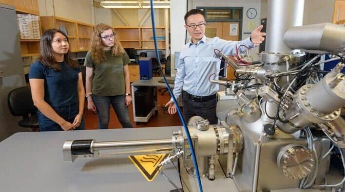
CAS: Chemistry & Biochemistry Dept: Surface Analysis Facility
DIRECTOR: Xu Feng, Ph.D.
ADDRESS: Lammot DuPont Laboratory, Newark, DE 19716
CONTACT: Surface Analysis Facility
LOCATED IN: Lammot DuPont Laboratory
Core Facility OVERVIEW: The Surface Analysis Facility exists primarily to support the federally funded research projects of University of Delaware faculty members, and it has been subsidized from University sources to do so. To help defray operating costs, the Facility also welcomes, and the NSF encourages, collaborative interactions with other universities and colleges, as well as local, regional and national for-profit companies.
EQUIPMENT:
- IONTOF TOF.SIMS 5 system
- Thermo Scientific K-Alpha XPS system
- AFM-Raman
SERVICES:
- Time-of-Flight Secondary Ion Mass Spectrometry (TOF-SIMS)
- X-ray Photoelectron Spectroscopy (XPS or ESCA)
- Atomic Force Microscopy (AFM)
- Raman Spectroscopy
CAS: X-Ray Crystallography Laboratory Core Facility
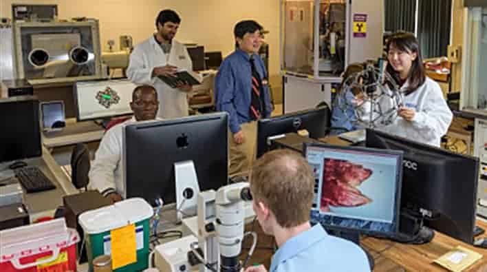
CAS: X-Ray Crystallography Laboratory Core Facility
ADDRESS: 236 Brown Laboratory, Newark, DE 19716
CONTACT: Glenn P. A. Yap, Ph.D.
LOCATED IN: Drake Hall/Brown Laboratory
Core Facility OVERVIEW: The X-ray Crystallography Laboratory conducts characterizations by X-ray diffraction of small-molecule organic or inorganic single crystals. As an extension service, the facility also accepts sample submissions from other departments of the University and from local, national and international collaborators of academe and industry. The facility also serves as an ancillary graduate research laboratory for selected graduate students pursuing studies in inorganic chemistry leading to a doctorate degree.
EQUIPMENT:
Dual wavelength APEX II Duo (Mo & Cu) Bruker-AXS CCD X-ray diffractometer. Data are collected typically at 200K. Structures are solved with SHELXTL software.
SERVICES:
Typical turnaround times under average conditions, from start of data-collection to completely refined structure, range from a few hours to a day.
CHS: Biostatistics Core Facility

CHS: Biostatistics Core Facility
DIRECTOR: Ryan Pohlig, Ph.D.
ADDRESS: 100 Discovery Blvd, STAR TOWER, 6th Floor, Newark, DE 19710
Core Facility OVERVIEW: The College of Health Sciences Biostatistics Core supports the development, conduct, and dissemination of research conducted in the College of Health Sciences and across the University of Delaware. In addition to CHS faculty researchers, we also support the development of student researcher through teaching graduate level courses student and mentorship. The work of the Biostatistics Core is critical for research progress in the College, which is ranked highly in terms of funding received from the National Institutes of Health for Schools of Allied Health. The Core supports a wide portfolio of externally-funded projects including R01s, R21s, the ACCEL-Center for Translational Research, and the Center for Biomedical Research Excellence in Cardiovascular Health (COBRE). Our statisticians are collaborators and an integral part of research teams and typically serve as funded personal on grant proposals. As such, they help shape the direction of research by providing methodological expertise that can improve science, build stronger teams, and lead to more competitive proposals for external funding.
What We Do – Building Research Partnerships
The core provides support in three main areas: collaboration around proposal development and research design; analysis and publication of data collected as part of funded research; and education. Click to request an opportunity for collaboration.
SERVICES:
- The Biostatistics Core partners with Principal Investigators on the development of Specific Aims, Research Questions, and Hypotheses; advises on research design; and provides power analyses and statistical analysis plans
- The Biostatistics Core performs data analyses, assists Principal Investigators in the interpretation of results, and contributes to the methods, results, and discussion sections of peer-reviewed abstracts and manuscripts.
- The Biostatistics Core teaches in the MPH in Epidemiology Program, offering courses to graduate students across the College of Health Sciences. In addition to formal courses, members of the Core may teach workshops and serve as mentors to individual graduate students, for example as members of a thesis or dissertation committee.
CARN: Food Sensory Characterization and Novel Processing Center (FSCNP)
CARN: Food Sensory Characterization and Novel Processing Center (FSCNP)
DIRECTOR: Dr Juzhong Tan, Ph.D.
ADDRESS: 529 S College Avenue, Newark, Delaware 19716
CONTACT: Dr. Juzhong Tan, Assistant Professor Sensory Science and Food Product Development
Core Facility and Center OVERVIEW: The Food Sensory, Characterization, and Novel Processing (FSCN) Center is a dynamic platform connecting the University of Delaware College of Agriculture and Natural Resources (CANR) with researchers and industry stakeholders. Our partners include food processors and manufacturers, startups, distributors, retailers, and technology companies.
Housed within the UD Department of Animal and Food Sciences, the FSCN Center delivers professional services, such as sensory evaluations, food processing, product characterization, and pilot-plant production across a diverse range of food products. We are also committed to fostering innovation by supporting the development and adoption of new food processing and analysis technologies through internal and external collaborations.
Our center has state-of-the-art laboratory and pilot-plant scale processing equipment, comprehensive analytical instruments, and a highly skilled team. We continually upgrade our facilities and technology to enhance our capacity for multidisciplinary research and industry partnerships.
For collaboration opportunities or to learn more about our services, please contact our center director, Dr. Juzhong Tan, at jztan@udel.edu.
Centers and Institutes
NOTE: Institutes & Centers reporting to Research Office 

